[vc_row][vc_column][vc_custom_heading text=»La vacuna para el síndrome del trocánter mayor» font_container=»tag:p|font_size:30|text_align:left|color:%231e1e1e» google_fonts=»font_family:Open%20Sans%3A300%2C300italic%2Cregular%2Citalic%2C600%2C600italic%2C700%2C700italic%2C800%2C800italic|font_style:600%20bold%20italic%3A600%3Aitalic»][ultimate_spacer height=»30″ height_on_tabs=»15″ height_on_tabs_portrait=»15″ height_on_mob_landscape=»15″ height_on_mob=»15″][/vc_column][/vc_row][vc_row][vc_column][vc_column_text]
Ya hemos expuesto en una entrada anterior (La epidemia de la cadera: EL Síndrome del Trocánter Mayor), que este síndrome tiene un nivel de incidencia muy elevada en la población de edad mayor, exixtiendo unacorrelación con la edad muy alta…. y llega a ser en algunos casos el factor principal de reducción de movilidad; ¡¿Y si estuviese en nuestras manos reducir el porcentaje de personas que caen en las garras de esta epidemia?! En una segunda entrada comprendimos que el mecanismo lesivo es el impingement externo de la cadera, donde la cintilla iliotibial comprime los tendones del glúteo medio y menor y las bursas sinoviales a la altura del trocánter mayor (Ver; Síndrome del Trocánter Mayor: La pescadilla que se muerde la cola).
El punto clave de la recuperación a largo plazo es tener una buena ratio Glúteo medio/Tensor de la fascia lata: Conforme aumentamos la fuerza de los abductores trocántereos, menos se requiere la participación de los abductores que tensan la cintilla iliotibial, por lo que disminuiremos el impingement externo, favoreciendo la regeneración de los tejidos dañados y su fortalecimiento, por lo que habremos invertido el sentido del círculo vicioso. Hay varios estudios electromiográficos que han buscado los ejercicios que mejoran esta ratio:
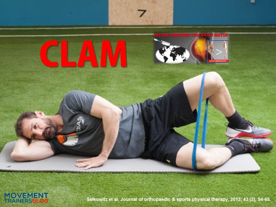
Selkowitz et al65 encontraron que el ejercicio que mejor valor obtenía de la ratio Glúteo medio+fibras superiores del glúteo mayor/Tensor de la Fascia Lata era el famoso CLAM, el problema es que es un ejercicio con unos niveles de activación baja de la musculatura abductora.
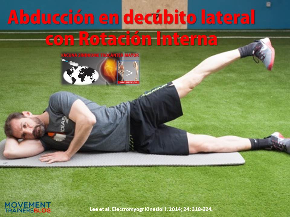
Lee et al45,46 con un escrupuloso control de la técnica del movimiento, encuentran mejores ratios de activación Gmed/TFL en la Abducción en decúbito lateral con la cadera en rotación interna (punta del pie hacia abajo) en una activación isométrica al 50% del ROM de abducción, al comparar el ejercicio con sus variantes en rotación externa o con cadera neutra.
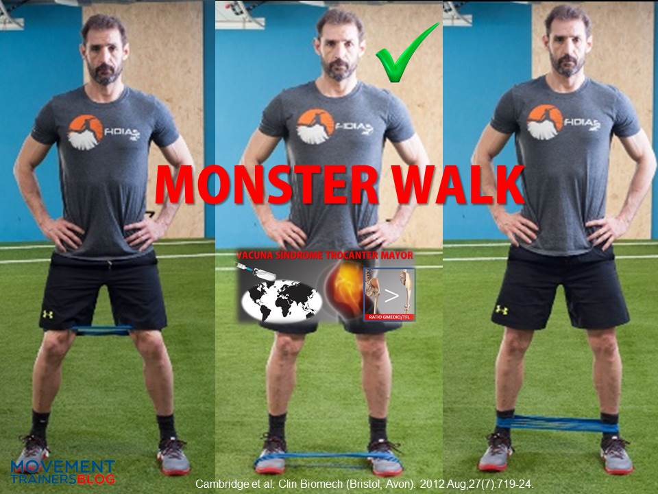
Y otros dos ejercicios que han demostrado tener una buena ratio Gmed/TFLson el pelvic drop en rotación interna54 (porque disminuye la eficiencia mecánica del tensor de la fascia lata al acortarlo) y el Monster Walk colocando la banda elástica en los pies13, esta banda exige una activación de la musculatura rotadora externa de la cadera inhibiendo los rotadores internos (TFL).

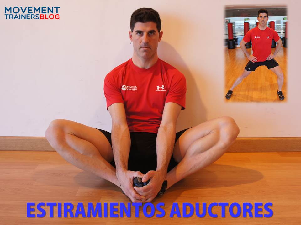
- Estiramientos de la cintilla iliotibial/abductores de cadera61. Se desaconseja en la fase aguda debido a que provoca el mecanismo lesivo del impingement externo, mientras que la disminución de su tensión, vía estiramientos puede ser de gran ayuda a medio/largo plazo, osea que lo utilizaremos más como trabajo preventivo que como ejercicio de recuperación.

- Evitar foam roller sobre el trocánter mayor en fases iniciales, ya que aumentamos la compresión transversal sobre los tejidos dañados mediante ese mismo mecanismo. Por el mismo motivo se desaconsejan posiciones de aducción de cadera como cruzar la piernas, o dormir de lado.
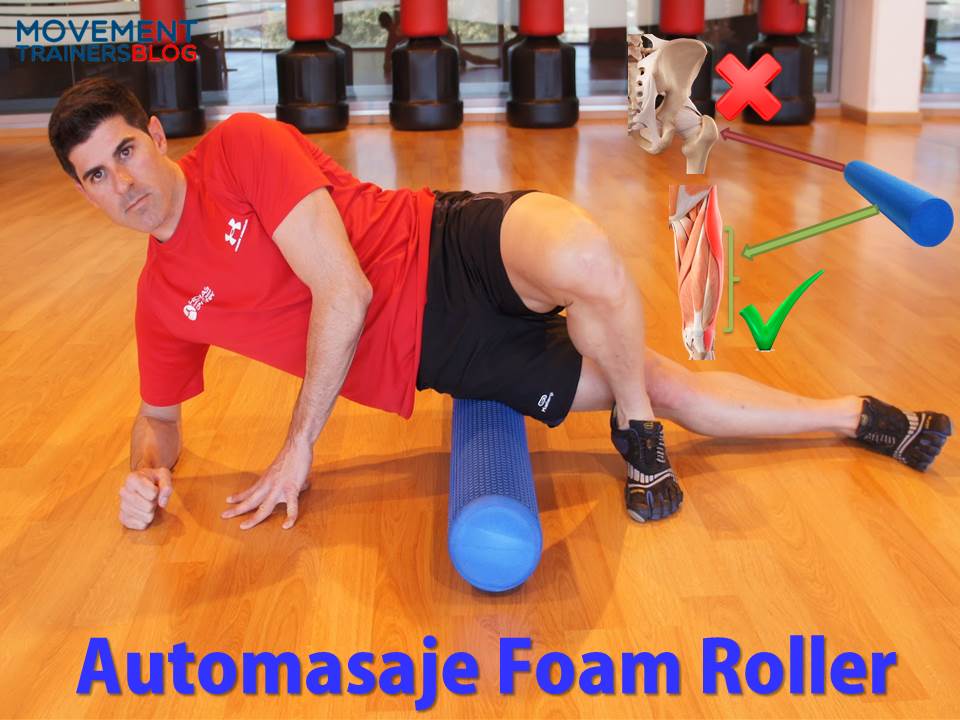
- Es importante disminuir la tensión de todos los músculos que tensan la cintilla iliotibial: TFL, fibras superiores del glúteo mayor y el vasto lateral. Por lo que sesiones de masaje o automasaje con el foam roller, o estiramientos en tensión activa o FNP de estos músculos van a reducir la tensión sobre la cintilla.
- Diversos autores55,59 recomiendan incluir trabajo excéntrico al igual que en otras tendinopatías, aunque no existen estudios concretos realizados para tendinopatías glúteas, pero a la espera de una confirmación científica, es lógico pensar que el comportamiento de estas tendinopatías puede ser similar ante el trabajo excéntrico.

- En corredores modificar la técnica de carrera si se hacen apoyos con excesiva aducción de cadera (ya que aumentaría el impingement externo), lo cual se puede mejorar con biofeedback visual56. Además de buscar una mayor cadencia17,36.
- Se recomienda un trabajo de fuerza (cargas longitudinales=tensión altas) tres veces por semana52. Un buen indicador de si las cargas son las adecuadas es la reducción del dolor nocturno.
- Aunque en caso de que una persona tenga un síndrome de aducción62, va a ser importante la ganancia de flexibilidad de la musculatura aductora.
Evidentemente no puede vivir una persona de ejercicios analíticos y fuera de su contexto deportivo o alejado de sus movimientos de la vida diaria, hay que buscar una progresión que permita trabajar los abductores de la cadera pero cada vez con tareas más específicas. Pero cuando hay un desequilibrio importante en la ratio GMed/TFL, es interesante centrarse un poco en la corrección estructural, aunque lo difícil será mejorar la ratio en gestos globales. Todos estos ejercicios tienen una sencilla forma de convertirse en tareas analíticas para sistemas complejos (Ver: Tuneando tareas analíticas), que es convirtiéndolos en juegos…… si los quieres conocer atento a la próxima entrada.
[ult_buttons btn_title=»¿TIENES ESTE PROBLEMA? SOLICITA UNA VALORACIÓN ONLINE GRATUITA | HAZ CLICK AQUÍ O LLAMA AL 956 872 619″ btn_link=»url:https%3A%2F%2Fcenter.fidias.net%2Fservicio-fidias-online%2F» btn_align=»ubtn-center» btn_size=»ubtn-large» btn_title_color=»#ffffff» btn_bg_color=»#d36200″ btn_bg_color_hover=»#e8e8e8″ btn_title_color_hover=»#d36200″ icon_size=»32″ btn_icon_pos=»ubtn-sep-icon-at-left» btn_font_family=»font_family:Montserrat|font_call:Montserrat|variant:regular» btn_font_style=»font-weight:normal;font-style:normal;» btn_font_size=»desktop:16px;»]
BIBLIOGRAFÍA:
-
Allison K, Bennell KL, Grimaldi A, Vicenzino B, Wrigley TV, Hodges PW. Single leg stance control in individuals with symptomatic gluteal tendinopathy. Gait Posture. 2016 Sep;49:108-13.
-
Allison K, Vicenzino B, Wrigley TV, Grimaldi A, Hodges PW, Bennell KL. Hip Abductor Muscle Weakness in Individuals with Gluteal Tendinopathy. Med Sci Sports Exerc. 2016 Mar;48(3):346-52.
-
Allison K, Wrigley TV, Vicenzino B, Bennell KL, Grimaldi A, Hodges PW. Kinematics and kinetics during walking in individuals with gluteal tendinopathy. Clin Biomech (Bristol, Avon). 2016 Feb;32:56-63.
-
Bancroft LW, Blankenbaker DG. Imaging of the tendons about the pelvis. AJR Am J Roentgenol. 2010;195:605-617.
-
Barnthouse NC, Wente TM, Voos JE. Greater trochanteric pain syndrome: endoscopic treatment options. Oper Tech Sports Med. 2012;20:320–324.
-
Berry JW, Lee TS, Foley HD, Lewis CL. Resisted Side Stepping: The Effect of Posture on Hip Abductor Muscle Activation. J Orthop Sports Phys Ther. 2015 Sep;45(9):675-82.
-
Bewyer DC, Bewyer KJ. Rationale for treatment of hip abductor pain syndrome. The Iowa Orthopaedic Journal. 2003;23:57-60.
-
Bird PA, Oakley SP, Shnier R, Kirkham BW. Prospective evaluation of magnetic resonance imaging and physical examination findings in patients with greater trochanteric pain syndrome. Arthritis Rheum. 2001 Sep;44(9):2138-45.
-
Birnbaum K, Siebert CH, Pandorf T, Schopphoff E, Prescher A, Niethard FU. Anatomical and biomechanical investigations of the iliotibial tract. Surgical and Radiologic Anatomy. 2004;26:433-46.
-
Blank E, Owens BD, Burks R, Belmont PJ Jr. Incidence of greater trochanteric pain syndrome in active duty US military service members. Orthopedics. 2012 Jul 1;35(7):e1022-7.
-
Brody, LT.; Hall, CM. Therapeutic exercise: Moving toward function. 3rd. Lippincott Williams & Wilkins; Baltimore: 2011.
-
Bunker TD, Esler CNA, Leach WJ. Rotator-Cuff Tear of the Hip. Journal of Bone & Joint Surgery – British Volume. 1997;79-B(4):618-20.
-
Cambridge ED, Sidorkewicz N, Ikeda DM, McGill SM. Progressive hip rehabilitation: The effects of resistance band placement on gluteal activation during two common exercises. Clin Biomech (Bristol, Avon). 2012; 27(7):719–24.
-
Chandrasekaran S, Vemula SP, Gui C, Suarez-Ahedo C, Lodhia P, Domb BG. Clinical Features That Predict the Need for Operative Intervention in Gluteus Medius Tears. Orthopaedic Journal of Sports Medicine. 2015;3(2):2325967115571079. doi:10.1177/2325967115571079.
-
Chi AS, Long SS, Zoga AC, Read PJ, Deely DM, Parker L, Morrison WB. Prevalence and pattern of gluteus medius and minimus endon pathology and muscle atrophy in older individuals using MRI. Skeletal Radiol. 2015 Dec;44(12):1727-33.
-
Chowdhury R, Naaseri S, Lee J, Rajeswaran G. Imaging and management of greater trochanteric pain syndrome. Postgrad Med J. 2014 Oct;90(1068):576-81.
-
Chumanov ES, Wille CM, Michalski MP, et al. Changes in muscle activation patterns when running step rate is increased. Gait Posture. 2012;36(2):231–5.
-
Clancy WG. Runners’ injuries: part two. Evaluation and treatment of specific injuries. Am J Sports Med. 1980;8(4):287–9.
-
Collee G. Greater trochanteric pain syndrome (trochanteric bursitis) in low back pain. Scand J Rheumatol. 1991;20:262–266
-
Cook JL, Purdam C. Is compressive load a factor in the development of tendinopathy? Br J Sports Med. 2012;46:163-168.
-
Cook JL, Purdam CR. The challenge of managing tendinopathy in competing athletes. Br J Sports Med. 2014;48:506-509.
-
Cook JL. Is tendon pathology a continuum? A pathology model to explain the clinical presentation of load-induced tendinopathy. British Journal of Sports Medicine. 2009;43(6):409-16.
-
Del Buono A, Papalia R, Khanduja V, et al. Management of the greater trochanteric pain syndrome: a systematic review. Br Med Bull. 2012;102:115–31.
-
Dwek J, Pfirrmann C, Stanley A, Pathria M, Chung CB. MR imaging of the hip abductors: normal anatomy and commonly encountered pathology at the greater trochanter. Magnetic Resonance Imaging Clinics Of North America. 2005;13(4):691.
-
Ebert JR, Retheesh T, Mutreja R, Janes GC. THE CLINICAL, FUNCTIONAL AND BIOMECHANICAL PRESENTATION OF PATIENTS WITH SYMPTOMATIC HIP ABDUCTOR TENDON TEARS. International Journal of Sports Physical Therapy. 2016;11(5):725-737.
-
Ege Rasmussen KJ, Fanø N. Trochanteric bursitis. Treatment by corticosteroid injection. Scand J Rheumatol. 1985;14(4):417-20.
-
Fearon AM, Cook JL, Scarvell JM, Neeman T, Cormick W, Smith PN. Greater Trochanteric Pain Syndrome Negatively Affects Work, Physical Activity and Quality of Life: A Case Control Study. J Arthroplasty. 2014;29(2):383-6.
-
Fearon AM, Stephens S, Cook JL, Smith PN, Cormick W, Scarvell JM. Are child bearing hips a risk factor for greater trochanteric pain syndrome? J Bodyw Mov Ther. 2012;16:148.
-
Fredericson M, Cookingham CL, Chaudhari AM, Dowdell BC, Oestreicher N, Sahrmann SA. Hip abductor weakness in distance runners with iliotibial band syndrome. Clin J Sport Med. 2000; 10(3):169–75
-
Furia JP, Rompe JD, Maffulli N. Low-energy extracorporeal shock wave therapy as a treatment for greater trochanteric pain syndrome. The American journal of sports medicine. 2009;37(9):1806- 1813.
-
Gaida JE, Ashe MC, Bass SL, Cook JL. Is adiposity an underrecognized risk factor for tendinopathy? A systematic review. Arthritis Rheum. 2009;61(6):840–9.
-
Grimaldi A, Fearon A. Gluteal Tendinopathy: Integrating Pathomechanics and Clinical Features in Its Management. J Orthop Sports Phys Ther 2015;45(11):910-922.
-
Grimaldi A, Mellor R, Hodges P, Bennell K, Wajswelner H, Vicenzino B. Gluteal Tendinopathy: A Review of Mechanisms, Assessment and Management. Sports Med. 2015;48(8):1107-19.
-
Grimaldi A. Assessing lateral stability of the hip and pelvis. Manual Therapy. 2011;16:26-32.
-
Grimaldi A, Richardson C, Stanton W, et al. The association between degenerative hip joint pathology and size of the gluteus medius, gluteus minimus and piriformis muscles. Man Ther. 2009;14(6):605–10.
-
Heiderscheit BC. Lower extremity injuries: is it just about hip strength? J Orthop Sports Phys Ther. 2010;40(2):39–41.
-
Ho GW, Howard TM. Greater trochanteric pain syndrome: more than bursitis and iliotibial traction friction. Curr Sports Med Rep. 2012 Sep-Oct;11(5):232-8.
-
Huang BK, Campos JC, Michael Peschka PG, Pretterklieber ML, Skaf AY, Chung CB, Pathria MN. Injury of the gluteal aponeurotic fascia and proximal iliotibial band: anatomy, pathologic conditions, and MR imaging. Radiographics. 2013 Sep-Oct;33(5):1437-52.
-
Iorio R, Healy WL, Warren PD, Appleby D. Lateral trochanteric pain following primary total hip arthroplasty. J Arthroplasty. 2006;21:233-236.
-
Kagan II A. Rotator cuff tears of the hip. Clin Orthop. 1999;368: 135–40.
-
Kagan II A. Rotator-cuff tear of the hip. J Bone Joint Surg (Br). 1998;80(1):182–3.
-
Kaltenborn A, Bourg CM, Gutzeit A, Kalberer F. The Hip Lag Sign – Prospective Blinded Trial of a New Clinical Sign to Predict Hip Abductor Damage. PLoS One. 2014; 9(3): e91560.
-
Ker RF, Wang XT, Pike AV. Fatigue quality of mammalian tendons. The Journal of experimental biology. 2000;203(Pt 8):1317-27.
-
Kimpel DM, Garner CC, Magone KM, May JH, Lawless MW. Greater trochanteric hip 583 pain. Orthop Nurs. 2014 Mar-Apr;33(2):95-9.
-
Lee J, Cynn H, Choi S, Yoon T & Jeong H. (2013). Effects of Different Hip Rotations on Gluteus Medius and Tensor Fasciae Latae Muscle Activity During Isometric Side-Lying Hip Abduction. Journal of Sport Rehabilitation, 22, 301-307.
-
Lee JH, Cynn HS, Kwon OY, et al. Different rotations hip abductor muscles activity influence during isometric side-lying hip abduction in subjects with gluteus medius weakness. Electromyogr Kinesiol J. 2014; 24: 318-324.
-
Leonard MH. Trochanteric syndrome; calcareous and noncalcareous tendonitis and bursitis about the trochanter major. J. Am. Med. Assoc. 1958; 168:175Y7
-
Lequesne M, Mathieu P, Vuillemin-Bodaghi V, Bard H, Djian P. Gluteal tendinopathy in refractory greater trochanter pain syndrome: diagnostic value of two clinical tests. Arthritis Rheum. 2008;59:241-246.
-
Lindner D, Shohat N, Botser I, Agar G, Domb BG. Clinical presentation and imaging results of patients with symptomatic gluteus medius tears. Journal of Hip Preservation Surgery. 2015;2(3):310-315.
-
Long SS, Surrey DE, Nazarian LN. Sonography of greater trochanteric pain syndrome and the rarity of primary bursitis. AJR Am J Roentgenol. 2013;201:1083-1086.
-
Magnusson SP NM, Maganaris CN et al. Human tendon behaviour and adaptation, in vivo. J Physiol. 2008;30(1616-20).
-
Magnusson SP, Langberg H, Kjaer M. The pathogenesis of tendinopathy: balancing the response to loading. Nat Rev Rheumatol. 2010;6:262-268.
-
Mellor R, Grimaldi A, Wajswelner H, Hodges P, Abbott JH, Bennell K, Vicenzino B. Exercise and load modification versus corticosteroid injection versus ‘wait and see’ for persistent gluteus medius/minimus tendinopathy (the LEAP trial): a protocol for a randomised clinical trial. BMC Musculoskelet Disord. 2016 Apr 30;17:196.
-
Monteiro RL, Facchini JH, de Freitas DG, Callegaric B, Amado João SM. Hip Rotations Influence Electromyographic Activity of Gluteus Medius Muscle During Pelvic Drop Exercise. J Sport Rehabil. 2016 Aug 24:1-21.
-
Mulligan EP, Middleton EF, Brunette M. Evaluation and management of greater trochanter pain syndrome. Phys Ther Sport. 2015 Aug;16(3):205-14.
-
Noehren B, Scholz J, Davis I. The effect of real-time gait retraining on hip kinematics, pain and function in subjects with patellofemoral pain syndrome. Br J Sports Med. 2011;45(9):691–6.
-
Pfirrmann CW, Chung CB, Theumann NH, et al. Greater trochanter of the hip: attachment of the abductor mechanism and a complex of three bursae—MR imaging and MR bursography in cadavers and MR imaging in asymptomatic volunteers. Radiology. 2001;221(2): 469–77.
-
Pfirrmann CW, Notzli HP, Dora C, et al. Abductor tendons and muscles assessed at MR imaging after total hip arthroplasty in asymptomatic and symptomatic patients. Radiology. 2005;235(3):969–76.
-
Reid D. The management of greater trochanteric pain syndrome: A systematic literature review. Journal of Orthopaedics. 2016;13(1):15-28. doi:10.1016/j.jor.2015.12.006.
-
Robertson WJ, Gardner MJ, Barker JU, Boraiah S, Lorich DG, Kelly BT. Anatomy and dimensions of the gluteus medius tendon insertion. Arthroscopy. 2008;24:130-136.
-
Rompe JD et al. Home training, local corticosteroid injection, or radial shock wave therapy for greater trochanter pain syndrome. Am J Sports Med. 2009; 37(10):1981–90.
-
Sahrmann, SA. Diagnosis and treatment of movement impairment syndromes. Mosby, Inc; St. Louis, MO: 2002.
-
Selkowitz, Beneck & Powers (2013). Which Exercises Target the Gluteal Muscles While Minimizing Activation of the Tensor Fascia Lata? Electromyographic Assessment Using Fine-Wire Electrodes. Journal of orthopaedic & sports physical therapy. 43 (2), 54-65
-
Schapira D, Nahir M, Scharf Y. Trochanteric bursitis: a common clinical problem. Arch Phys Med Rehabil. 1986 Nov;67(11):815-7.
-
Selkowitz DM, Beneck GJ, Powers CM. Which exercises target the gluteal muscles while minimizing activation of the tensor fascia lata? electromyographic assessment using fine-wire electrodes. J Orthop Sports Phys Ther. 2013; 43(2):54–64.
-
Shbeeb MI, Matteson EL. Trochanteric bursitis (greater trochanter pain syndrome). Mayo Clin Proc. 1996 Jun;71(6):565-9.
-
Sim FH, Scott SG. Injuries of the pelvis and hip in athletes: anatomy and function. In: Nicholas JA, Hershman EB, eds. The lower extremity and spine in sports medicine. St Louis, Mo: Mosby; 1986:1119-1169.
-
Stecco A, Gilliar W, Hill R, et al. The anatomical and functional relation between gluteus maximus and fascia lata. J Bodyw Mov Ther. 2013;17(4):512–7.
-
Stegemann H. Die chirurgische bedevtung paraartikularer kalkablagerungen. Arch. Klin. Chir. 1923; 125:718Y38.
-
Stern JR. Anatomical and functional specializations of the human gluteus maximus. Am J Phys Anthrop. 1972;36:315–40.
-
Strauss EJ, Nho SJ, Kelly BT. Greater trochanteric pain syndrome. Sports Med Arthrosc 2010;18: 113–119.
-
Sutter R, Kalberer F, Binkert CA, et al. Abductor tendon tears are associated with hypertrophy of the tensor fasciae latae muscle. Skel Radiol. 2013;42(5):627–33.
-
Thornton GM, Shao X, Chung M, et al. Changes in mechanical loading lead to tendon-specific alterations in MMP and tIMP expression: influence of stress deprivation and intermittent cyclic hydrostatic compression on rat supraspinatus and Achilles tendons. Br J Sports Med. 2010;44(10):698–703.
-
Vleeming A, Mooney V, Snijders C, et al. Movement, stability and low back pain: the essential role of the pelvis. New York: Churchill Livingstone; 1997.
-
Walsh G. MRI in greater trochanter pain syndrome. Australas Radiol. 2003;47(1):85–7.
-
Williams BS, Cohen SP. Greater trochanteric pain syndrome: a review of anatomy, diagnosis and treatment. Anesth Analg 2009;108:1662–70.
-
Williams RL, Warwick R. Gray’s Anatomy. 36th ed. Philadelphia, PA: Saunders; 1980.
-
Winston P, Awan R, Cassidy JD, et al. Clinical examination and ultrasound of self-reported snapping hip syndrome in elite ballet dancers. Am J Sports Med. 2007;35:118–126.
-
Woodley SJ, Mercer SR, Nicholson HD. Morphology of the bursae associated with the greater trochanter of the femur. J Bone Joint Surg Am 2008;90:284–94
-
Woodley SJ, Nicholson HD, Livingstone V, et al. Lateral hip pain: findings from magnetic 789 resonance imaging and clinical examination. J Orthop Sports Phys Ther. 2008;38:313-328.
-
Woyski D, Olinger A, Wright B. Smaller insertion area and inefficient mechanics of the 791 gluteus medius in females. Surg Radiol Anat. 2013;35:713-719.
-
Yen YM, Lewis CL, Kim YJ. Understanding and Treating the Snapping Hip. Sports Med Arthrosc. 2015 Dec;23(4):194-9.
-
Youdas JW, Foley BM, Kruger BL, et al. Electromyographic analysis of trunk and hip muscles during resisted lateral band walking. Physiother Theory Pract. 2013; 29(2):113–23.
[/vc_column_text][/vc_column][/vc_row][vc_row][vc_column][ultimate_spacer height=»20″ height_on_tabs=»10″ height_on_tabs_portrait=»10″ height_on_mob_landscape=»10″ height_on_mob=»10″][ult_buttons btn_title=»¿TIENES ESTE PROBLEMA? SOLICITA UNA VALORACIÓN ONLINE GRATUITA | HAZ CLICK AQUÍ O LLAMA AL 956 872 619″ btn_link=»url:https%3A%2F%2Fcenter.fidias.net%2Fservicio-fidias-online%2F» btn_align=»ubtn-center» btn_size=»ubtn-large» btn_title_color=»#ffffff» btn_bg_color=»#d36200″ btn_bg_color_hover=»#e8e8e8″ btn_title_color_hover=»#d36200″ icon_size=»32″ btn_icon_pos=»ubtn-sep-icon-at-left» btn_font_family=»font_family:Montserrat|font_call:Montserrat|variant:regular» btn_font_style=»font-weight:normal;font-style:normal;» btn_font_size=»desktop:16px;»][vc_custom_heading text=»CURSOS RELACIONADOS» font_container=»tag:p|font_size:22|text_align:left|color:%23d36200″ google_fonts=»font_family:Open%20Sans%3A300%2C300italic%2Cregular%2Citalic%2C600%2C600italic%2C700%2C700italic%2C800%2C800italic|font_style:600%20bold%20italic%3A600%3Aitalic»][ultimate_spacer height=»20″ height_on_tabs=»10″ height_on_tabs_portrait=»10″ height_on_mob_landscape=»10″ height_on_mob=»10″][dt_portfolio_carousel dis_posts_total=»» posts_offset=»0″ content_alignment=»center» image_sizing=»proportional» image_border_radius=»3px» image_scale_animation_on_hover=»disabled» image_hover_bg_color=»disabled» slides_on_wide_desk=»3″ item_space=»20″ link_lead=»follow_link» post_date=»n» post_category=»n» post_author=»n» post_comments=»n» post_content=»off» read_more_button=»off» show_link=»n» show_zoom=»n» show_details=»n» project_icon_border_width=»0px» project_icon_color=»#ffffff» project_icon_color_hover=»#ffffff» arrow_bg_width=»36x» arrow_border_width=»0px» r_arrow_icon_paddings=»0px 0px 0px 0px» r_arrow_v_offset=»0px» l_arrow_icon_paddings=»0px 0px 0px 0px» l_arrow_v_offset=»0px» category=»1286, 2160″][vc_separator border_width=»2″][/vc_column][/vc_row]


